ABSTRACT
Renal calculi, commonly known as kidney stones, present a significant health concern worldwide, affecting millions of individuals annually. Conventional treatments often include medication, lithotripsy, or surgical intervention, yet these approaches may carry risks and side effects. Homeopathy offers a holistic alternative that aims to not only alleviate symptoms but also address the underlying causes of renal calculi formation. This article provides an overview of the pathophysiology, symptoms, and conventional treatments of renal calculi, followed by a detailed exploration of the principles and practice of homeopathy in managing this condition. Drawing upon clinical evidence and case studies, we highlight key homeopathic remedies commonly prescribed for renal calculi, along with their corresponding symptomatology and constitutional indications. Furthermore, we discuss the importance of individualized treatment plans and the potential benefits of adjunctive lifestyle modifications and dietary measures in preventing recurrence. By integrating homeopathy into the therapeutic landscape of renal calculi management, patients can potentially experience improved symptom relief, enhanced overall well-being, and reduced reliance on conventional interventions.
KEYWORDS
Renal calculi, Kidney stones, Nephrolithiasis, urolithiasis, homoeopathy.
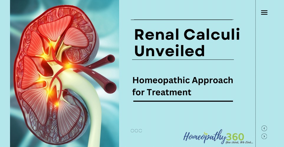
RENAL CALCULI
INTRODUCTION
- RENAL CALCULI ALSO KNOWN AS UROLITHIASIS, IS A KIDENY STONE DISEASE WHERE A SOLID PIECE OF MATERIAL (KIDNEY STONE) OCCURS IN THE URINARY TRACT.
- A kidney stone is a hard solid mass of material that forms in the kidney from the substances in the urine.
- Kidney stones develop as a result of various metabolic disorders which affect the fate of calcium and other mineral elements in the body.
- Stones may be formed in the kidney , urinary bladder, ureter and urethra.
INCIDENCE
- Urinary calculi are more common in men than in women
- Incidence of urinary calculi peaks between the 3rd and 5th decades of life.
- 80% of stones are under 2mm in size
- 90% of stones pass through the urinary system spontaneously
- There is seasonal variation with stone occurring more often in the summer months suspecting the role of dehydration in this process.
ETIOLOGY
SUPERSATURATION OF URINE
- Crystals when in a supersaturated concentration, can precipitate and unite to form a stone.
- Keeping urine dilute and free flowing reduces the risk of recurrent stone formation in many individuals.
- It is known that a mucoprotein is formed (the matrix for the stone) in the kidneys that forms stones.
OBSTRUCTION WITH URINARY STASIS
URINARY PH-
- The higher the PH (alkaline) the less soluble are calcium and phosphate.
- The lower the PH (acidic) the less soluble are uric acid and cystine.
LACK OF INHIBITORS
- Lack of inhibitors increase the risk of stone formation
- Inhibitor substances such as citrate and magnesium appear to keep particles from aggregating and forming crystals.
CERTAIN MEDICATIONS-
Eg-
- Calcium carbonate
- Sodium bicarbonate
- Aluminium hydroxide
RISK FACTORS
Immobility and sedentary lifestyle, obesity, dehydration, which leads to super saturation- Low fluid intake that lead to increased urinary concentration.
CLIMATE
- Warm climates that causes increased fluid loss
- low urine volume
- Increased solute concentration in urine.
PREVIOUS HISTORY OF RENAL CALCULI
DIETARY INTAKE
- A diet rich in purine, oxalates, calcium supplements, animal proteins
- Excessive amounts of tea or fruit juices that elevate urinary oxalate level.
HIGH MINERAL CONTENT IN DRINKING WATER
GOUT
GENETIC FACTORS- Family history of stones formation, cystinuria, gout or renal acidosis.
PROLONGED INDWELLING CATHETERIZATION
NEUROGENIC BLADDER (FLACCID OS SPASTIC)
TYPES OF RENAL CALCULUS
The stones may be of one crystal type or combination of types.
- CALCIUM OXALATE
- CALCIUM PHOSPHATE
- STRUVITE
- URIC ACID
- CYSTINE
- XANTHINE
CALCIUM OXALATE
- This type of stone is usually single and is extremely hard .
- It is dark in color due to the staining with altered blood precipitated on its surface.
- Spiky – covered with sharp projections – which causes bleeding due to injury to the adjacent tissues.
- MULBERRY STONE
PECULIARITIES OF CALCIUM OXALATE
- It is often impacted in the ureter
- It causes bleeding due to its rough surface
- Deposits of secondary phosphates on its surface leads to mixed stone
- Due to high calcium content, it casts an exceptionally good shadow radiologically (radiopaque)
- Rough surface may also be evident in x-ray
URIC ACID CALCULI
- Pure uric acid stones are rare.
- Sensitivity – radiolucent – Not visible on x-ray (non radio-opaque)
- These stones usually occur multiple
- Moderate hardness
- Colour – yellow to dark brown
- These stones usually occur in acidic urine
CYSTINE
- Cystine is an amino acid rich in sulphur Usually occur in multiple
- Colour – soft yellow/pink in colour
- When these are exposed outside the hue gradually changes to green
- Sensitivity – pure cystine stones are non radio-opaque but as they contain sulphur they are radio-opaque
- Such stones also occur in acid urine
CALCIUM PHOSPHATE
- Colour – dirty white
- Sensitivity – radio opaque
- Mixed stone (typically) with struvite or oxalate stones
STRUVITE CALCULI (MAGNESIUM AMMONIUM PHOSPHATE)
- Known as triple phosphate
- Colour – dirty white in color
- Smooth, soft and friable
- This type of calculus usually occurs in infected urine and so-called secondary calculus.
- Such stone enlarges rapidly and gradually fills up pelvis and renal calyces to take up the shape of Staghorn calculus.
STAGHORN CALCULUS
- Sensitivity – radio opaque
XANTHINE CALCULI
- Extremely rare
- Colour – brick red in colour, smooth round
PATHOPHYSIOLOGY
Calcium and oxalate come together to make the crystal nucleus
Supersaturation promotes their combination (as does inhibition)
Continued deposition at the renal papillae leads to growth of the kidney stone
Kidney stone grow and collect debris
In the Case where the kidney stones block all routes to the renal papillae, this can cause severe discomfort.
STAGES OF STONE FORMATION
SATURATION-SUPERSATURATION-NUCLEATION-CRYSTALLISATION-AGGREGATION-CRYSTAL RETENTION- STONE FORMATION
CLINICAL MANIFESTATIONS
The most characteristic manifestation of renal or ureteric calculi is a sharp, severe pain of sudden onset, caused by movement of calculus and consequent irritation.
(depending on the site of the stone, the pain may be either renal colic or ureteral colic).
SYMPTOM WISE CAN BE DIVIDED INTO
4 GROUPS
- QUIESCENT CALCULUS
- PAIN
- HYDRONEPHROSIS
- HAEMATURIA
1.QUIESCENT CALCULUS
- A few stones, particularly the phosphate stones, may lie dormant for quite long period.
- During this time the stones gradually increase in size with destruction of parenchyma
- Such stones may be discovered accidentally in x-ray performed for some other resons.
2. PAIN –
A) RENAL COLIC-
- originates deep in the lumbar region and radiates around the side and down towards the testicle in the male and the bladder in the female.
- If the stone is free and obstructs a calyx or ureteropelvic junction, there will be dull flank pain.
- The pain is situated in the renal angle posteriorly and in the corresponding hypochondrium anteriorly.
- The pain gets worse on movement and going upstairs.
B) URETERIC COLIC-
- This occurs when the stone attempts to pass down the ureter or temporarily blocks the pelviureteric junction.
- Agonising pain, which radiated from loin to groin.
- Pain comes on suddenly during which the patient rolls about drawing up his knees towards the chest, tossing on the bed in agony.
- When the pain is severe there will be profuse, sweating, nausea, vomiting, pallor, grunting respiration, elevated BP and PR, anxiety.
C. RADIATION OF PAIN
- The typical radiation of colicky pain is due to reflex pain which takes place along the course of Iliohypogastric and ilioinguinal nerves, which are the somatic nerves of the same segments which supply the autonomic nervous system to ureter (T11, 12 & L1)
- When the stone is in the lower part of the ureter the pain is referred to the scrotum or labia majora and inner side of the thighs along the distribution of the genitofemoral nerve.
- When the stone is in the intramural part of ureter, strangury may occur.
- STRANGURY MEANS – painful straining at urination in vain, with passage of few drops of blood stained urine.
D. REFERRED PAIN
- This is quite rare and is sometimes referred to all over the abdomen.
- Such pain may mimic peptic ulcer or gallbladder disease.
- Sometimes pain may be referred to opposite kidney which is called – RENORENAL REFLEX
3. HYDRONEPHROSIS
- Sometimes patients may complain of a lump in the loin and dull ache which are due to hydronephrosis due to renal stone.
4.HAEMATURIA
- Occasionally haematuria is the leading and only symptom.
OTHERS-
- Infection with elevated temperature and WBC count and urine obstruction that causes hydroureter, hydronephrosis or both.
PHYSICAL SIGNS
TENDERNESS –
- This is mostly present in the renal angle posteriorly between lower border of 12th rib and lateral border of erector spinae muscles – COSTOVERTEBRAL ANGLE.
- Anteriorly such tenderness may be elicited about an inch below and medial to the tip of 9th costal cartilage, which is known as RENAL POINT.
- MUSCLE RIGIDITY – over the kidney in few cases.
- SWELLING – where there is hydronephrosis or pyonephrosis associated with renal calculus, a swelling may be felt in the flank.
INVESTIGATIONS
BLOOD –
- ESR
- S.CALCIUM, PHOSPHATE, CREATININE, BLOOD UREA, URIC ACID
URINE
- Calcium, urate, cystine if suspected only
- PH, specific gravity
- Urine analysis
- c/s to rule out bacterial infection
PLAIN X-RAY , KUB
- To see kidney shadow stones
- (90% – radio opaque)
USG-ABDOMEN
- Can detect radiolucent stones and gives information about the changes in renal parenchyma.
- It is less useful when attempting to locate stones trapped in the midureter
CT-
- WILL IDENTIFY THE SMALL MISSED STONES IN URETER.
- To differentiate non opaque stone from a tumour.
COMPLICATIONS
- Infection and sepsis
- Obstruction of urinary tract
- Hydronephrosis
- Hydroureter
- Renal failure
- Renal haematoma
- Hypertension
MANAGEMENT
CONSERVATIVE-
- To relieve the pain until its causes can be eliminated.
- To eradicate the stone
- To determine the stone type
- To prevent nephron destruction
- To control infection
- To relieve any obstruction
INCREASE FLUID INTAKE
- To facilitate passage of small stones and to prevent development of new ones.
- To increase fluids 3-4 L daily to ensure urine output 2.5 to 3L daily
- Decreases the concentration of solutes and alleviates urinary stasis
PREVENT STONE RECURRENCE
- Diet modifications and medications
SURGICAL MANAGEMENT
OBSTRUCTION RELIEF-
- Ureteral stent insertion- Double J Stent
- PERCUTANEOUS NEPHROSTOMY- Placement of small, flexible tubes through skin into the kidney to drain urine.
- It is inserted through the back or flank.
DEFINITE SURGICAL TREATMENT-
- EXTRACORPOREAL SHOCK-WAVE LITHOTRIPSY
- LASER LITHOTRIPSY
- PERCUTANEOUS NEPHROLITHOTOMY
- URETEROSCOPY
- PERCUTANEOUS STONE DISSOLUTION
- CYSTOLITHOTOMY
- PARTIAL TOTAL NEPHRECTOMY
ESWL–
- This method uses ULTRASONIC WAVES TO BREAK UP STONES.
- Then the stones leave the body in the urine.
LASER LITHOTRIPSY
- Laser is used to break stones in to pieces.
PERCUTANEOUS NEPHROLITHOTOMY
- Used for large stones in or near the kidney or when kidneys or surrounding area are incorrectly formed.
- The stone is removed with an endoscope that is inserted into the kidney through small opening.
URETEROSCOPY WITH LASER LITHOTRIPSY
- Used for stones in the lower urinary tract.
- It involves first visualizing the stone and then destroying it.
- Inserting an ureteroscope into ureter and then inserting a laser electrohydraulic lithotripter or ultrasound device through ureteroscope to fragment and remove stone.
PERCUTANEOUS STONE DISSOLUTION
- Using infusions of chemical solutions (chemo lysis) such as alkylating agents, acidifying agents for the purpose of dissolving the stone.
CYSTOLITHOTOMY
- Removal of bladder calculi through a suprapubic incision is used only stones cannot be crushed and removed transurethral.
PARTIAL/TOTAL NEPHRECTOMY
- Is necessary because of extensive kidney damage, overwhelming renal infection, abnormal renal parenchyma, which can be responsible for stone formation.
BENCH SURGERY
- Kidney is removed out, temporarily.
HOMOEOPATHIC MANAGEMENT STRATEGIES FOR RENAL CALCULI
Homeopathic therapeutics for renal calculi aim to address the symptoms associated with kidney stones and promote the body’s natural ability to eliminate them. Here’s how homeopathy approaches the treatment of renal calculi:
INDIVIDUALIZATION: Homeopathy emphasizes the importance of individualizing treatment based on the unique symptoms and characteristics of each patient. A homeopath will conduct a detailed assessment of the patient’s symptoms, medical history, lifestyle, and emotional state to select the most appropriate remedy.
SYMPTOM SIMILARITY: Homeopathy operates on the principle of “like cures like,” which means that a substance capable of producing symptoms in a healthy person can be used to treat similar symptoms in a diseased person when administered in a highly diluted form. Homeopathic remedies for renal calculi are selected based on their ability to mimic the symptoms experienced by the patient, such as sharp, cutting pains in the back or abdomen, urinary urgency, burning during urination, and blood in the urine.
CONSTITUTIONAL TREATMENT: Homeopathy considers the patient as a whole, addressing not only the physical symptoms but also the mental, emotional, and constitutional aspects of health. A constitutional remedy is chosen based on the patient’s overall temperament, personality traits, and other individual characteristics. This holistic approach aims to strengthen the body’s inherent healing mechanisms and address the underlying imbalances that may contribute to kidney stone formation.
SELECTION OF REMEDIES: There are several homeopathic remedies commonly used in the management of renal calculi, each with its own unique symptom profile and indications. Some of the commonly prescribed remedies include Berberis vulgaris, Lycopodium clavatum, Cantharis vesicatoria, Sarsaparilla officinalis, and Calcarea carbonica. The choice of remedy depends on the specific symptoms experienced by the patient, as well as their individual constitution.
ADJUNCTIVE MEASURES: In addition to prescribing homeopathic remedies, homeopaths may recommend lifestyle modifications and dietary changes to support kidney health and prevent the recurrence of kidney stones. This may include increasing fluid intake, reducing the consumption of foods that can contribute to stone formation (such as those high in oxalates or purines), and adopting a balanced diet rich in fruits, vegetables, and whole grains.
Overall, homeopathic therapeutics for renal calculi offer a holistic and individualized approach to managing the symptoms of kidney stones, promoting overall health, and reducing the risk of recurrence through natural, non-invasive treatment strategies.
HOMOEOPATHIC THERAPEUTICS
SARASAPARILLA
- Severe, almost unbearable pain at conclusion of urination.
- Passage of gravel or small calculi; renal colic; stone in bladder; bloody urine.
- Urine: bright and clear but irritating; scanty, slimy, flaky, sandy, copious, passed without sensation, deposits white sand.
- Painful distention and tenderness in bladder; urine dribbles while sitting, standing, passes freely; air passes from urethra.
MANGANUM ACETICUM
- Biliary calculi & renal calculi
- Frequent urging to urinate.
- Cutting in middle of urethra between micturition.
- Urine profuse ; sediment violet, earthy.
BERBERIS VULGARIS
- Remedy for renal colic, especially when pain in left side extended from kidney region to urethra, with intense urging to urinate.
- Berberis vulgaris helps in soothing the pain in the lower back or the pain that shoots towards the bladder.
- It also helps in reducing the bubbling sensation and relieves the other discomforts caused due to kidney stones.
- Similar symptoms in right side also be cured.
HYDRANGEA
- Hydrangea is famous as the ‘stone breaker’.
- It breaks kidney stones in the ureter and urinary bladder.
- This remedy is indicated for gravel and deposition of white amorphous salts in urine.
- Burning in urethra with frequent desire to urinate.
- Renal calculi with a sharp pain in loins especially left side.
- Urine hard to start.
- Heavy deposit of mucus.
- Sharp pain in loins, especially left.
- Spasmodic stricture.
PAREIRA BRAVA
- The urinary symptoms are most important.
- Useful in renal colic, prostatic affections, and catarrh of bladder.
- Sensation as if bladder were distended, with pain.
- Pain going down thigh.
- The constant urging of urination with great straining.
- A person can pass urine only when he sits on his knees, pressing head firmly against the floor.
TEREBINTHINA
- Violent drawing pain in the region of
- kidneys. Violent burning and cutting pain during urethra.
- Blood and albumin in urine.
- It is said to prevent and dissolve renal calculi.
URTICA URENS
- Among the homeopathic treatments for kidney stones with high uric acid levels, this one is particularly effective (gout).
- Urtica urens should be considered in all cases of kidney stones involving a high uric acid levels.
- In such instances, Urtica urens is the best homeopathic remedy for kidney stones since, it removes the stone effectively.
SOLIDAGO
- Scanty, reddish-brown, thick sediment, dysuria,gravel.
- Difficult and scanty.
- Albumen, blood and slime in urine.
- Pain in kidneys extends forward to the abdomen and bladder.
- Clear and offensive urine.
- Sometimes makes the use of the catheter unnecessary.
UVA URSI
- Calculus inflammation.
- Chronic vesicle irritation with pain, tenesmus and catarrhal discharge.
- Burning after the discharge of slimy urine.
- Frequent urging with severe spasms of the bladder.
- Urine contains blood, pus and much tenacious mucous with clots in large masses.
- Painful dysuria.
- Involuntary green urine.
- Cystitis with bloody urine.
CANTHARIS
- Constant and intolerable urging to urinate before, during and after urination.
- Burning, scalding urine with cutting, intolerable urging and fearful tenses or dribbling strangury.
- Urine is passed drop by drop.
- Intolerable urging with tenses.
- Urine scalds the passage.
- Jelly like shreddy urine.
BIBLIOGRAPHY
- Janson C. The Oxford Textbook of Surgery. The Yale Journal of Biology and Medicine. 1995 May;68(3-4):155.
- Doherty GM, Way LW, editors. Current diagnosis & treatment: surgery. New York, NY, USA: Lange Medical Books/McGraw-Hill; 2010
- Townsend CM, Beauchamp RD, Evers BM, Mattox KL, editors. Sabiston textbook of surgery: the biological basis of modern surgical practice. Elsevier Health Sciences; 2016 Apr 22.
- Bhat S. SRB’s Manual of Surgery. Jaypee Brothers Medical Publishers; 2019 Jun 30.
- Das S. A concise textbook of surgery. Dr. S. Das.; 2006.
- Nan AK. Undergraduate surgery. Academic Publishers; 1997.
- Kent JT. Lectures on homoeopathic materia medica. New Delhi, India:: Jain Publishing Company; 1980.
- Allen HC. Keynotes and Characteristics with Comparisons of some of the leading Remedies of the Materia Medica with Bowel Nosodes. B. Jain Publishers; 2002.
- Murphy R. Lotus Materia medica. B. Jain Publishers; 2003.
- Boericke W. Pocket manual of homoeopathic Materia Medica & Repertory: comprising of the characteristic and guiding symptoms of all remedies (clinical and pathogenetic [sic]) including Indian Drugs. B. Jain publishers; 2002.
- Nash EB. Leaders in homoeopathic therapeutics. Boericke & Tafel; 1913.
- Lilienthal S. Homoeopathic therapeutics. B. Jain Publishers; 1998.
- Clarke JH. A dictionary of practical Materia medica. homoeopathic publishing Company; 1902.
- Chakma A. Renal calculi: An evidence-based case study. Int J Med Allied Heal Sci. 2015 Aug;7(1):5-9.
- Suri MC. Scope of homoeopathy in treatment of renal calculi.


