Abstract:
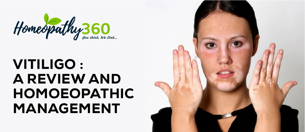
Vitiligo is a chronic disease characterised by loss of skin colour in patches because of disappearance of melanocytes in the affected area. This article deals with an overview of Vitiligo, including its etiological factors, pathogenesis, clinical patterns, clinical features and diagnosis along with homoeopathic management of the same, covering all three aspects namely organon, repertory and materia medica.
Keywords:
Vitiligo, autoimmune, genetic, homoeopathy.
Abbreviations:
Immunoglobulin (Ig), reactive oxygen species (ROS), hydrogen peroxide (H2O2)
Introduction: Vitiligo is a chronic systemic acquired disease that has an unpredictable clinical course, characterised by the appearance of macules and achromic or hypochromic patches on the skin and mucous membranes due to the disappearance of melanocytes in the affected area. These lesions can appear in different shapes and sizes.1Vitiligo has a pronounced impact on the physical and mental health of patients, including loss of skin photoprotection, compromised cutaneous immunity, and an appreciable reduction in quality of life2.The disease affects both genders equally, it can appear at any age. The prevalence of vitiligo has been estimated between 0.5% to 1.13% in India.1Vitiligo affects 1% of the general population, the risk of a patient’s sibling developing the disease is 6%, and for an identical twin 23% 3.
Site.–Vitiligo patches can appear anywhere on the skin, but commonly affected sites include the area around the orifices, the genitals or any sun-exposed areas such as the face and hands. The hair and rarely, the eyes may also be affected.4,5
Aetiology and precipitating factors-Exact aetiology is unknown and different patterns of vitiligo have different pathogenesis.4,5
- Family history- 20% patients have positive family history.
- Autoimmune hypothesis –association with other autoimmune disorder.
Presence of antibodies to melanocytes.
Presence to lymphocytes.
- Trauma.
Vitiligo pathogenesis-.
The pathogenesis is complex and involves the interplay of multiple factors. The pathogenesis of vitiligo elaborated by several theories like the neural hypothesis, autoimmune hypothesis and biochemical theory6.
Neural theory– Segmental vitiligo is present along a dermatome in distribution of nerve it has been hypothesised that a toxin which destroys melanocyte, is released at the nerve ending6.
The autoimmune hypothesis –According to this, vitiligo patients, immunoglobulin G (IgG) and immunoglobulin M (IgM) against melanocytes were found , low levels IgA may also be found 6.
Biochemical Theory- reactive oxygen species model-Oxidative stress hypothesis suggests that imbalanced redox (reduction-oxidation) state of the vitiliginous skin. This results in the dramatic production of reactive oxygen species (ROS), as H2O2. ROS oxidize components of the cell leading to melanocytes destruction and creating the depigmented macules6.
Patterns5–
- Segmental vitiligo.
- Non segmental vitiligo.
- Localised:
- Focal.
- Mucosal.
- Generalised:
- Vulgaris.
- Acrofacial.
- Universalis
- Segmental vitiligo-It occurs most commonly in children. Most commonly (50%) seen in distribution of trigeminal nerve(mandibular division). Leucotrichia on the depigmented area as well as away from vitiliginous areas frequent.
- Focal vitiligo- Acquired, small, 1-2 hypopigmented/depigmented lesion.
- Mucosal vitiligo– It occurs on mucous membranes (oral, lips, genital, both male and female).
- Vitiligo vulgaris- Common type of vitiligo. It occurs after 2nd decade, family history is present.
- Acrofacial vitiligo– vitiligo seen on face around eyes and on lips, ventral part of the hand and feet.
- Vitiligo universalis– widespread vitiligo with only few areas of normal pigmentation.
Clinical features5–
- Symmetrical, well defined, varied sized depigmented patches.
- Patchy loss of skin colour, which usually first appears on the hands, face, and areas around orifices.
- Premature whitening or graying of the hair on scalp, eyelashes, eyebrows or beard.
- Loss of colour in the tissues that line the inside of the mouth and nose (mucous membranes).
Diagnosis5– Diagnosis of vitiligo is usually clinical.
- Presence of depigmented macules (milky-white) with scalloped borders.
- Lecotrachia on affected area.
- Presence of Kobner’s phenomenon-New lesion of vitiligo at sites of trauma, appearing as linear depigmented macules.
Differential diagnosis5–
Nevus achromicus
- Pityriasis alba
- Leprosy.
- Tinea versicolor.
- Albinism.
HOMOEOPATHIC VIEWPOIN T
1. Miasmatic diagnosis- Complete absence or diminution of melanocytes from the birth is due to syphilitic miasm and cure by anti-syphilitic medicine. Case with the function of melanocytes is defective it represent sycotic miasm. Secondary depigmentation during the cause of illness due to psora, sycosis and syphilis or an combination of two or three of them.7,8,9
2. Repertorial approach– Vitiligo related rubrics are present in various commonly used repertories, which serves as a guide for selection of best simillimum.10,11,12,13
| Rubrics | Medicines | Repertory |
| SKIN – DISCOLORATION, – white | APIS ARS.calc. carb-v. Fl-ac. KALI-C.lac-c. sumb | Synthesis Repertory |
| SKIN – DISCOLORATION, – white – spots | Alum.am-c. ARS.Aur.Berb.Calc. carb-an. coca Merc.Nat-c.nit-ac. Phos. Sep. SIL. Sulph | Synthesis Repertory |
| SKIN – DISCOLORATION, – white | APIS ARS.calc. carb-v. Fl-ac. KALI-C.lac-c. sumb | Kent Repertory |
| Skin – VITILIGO- | sep.thuj. | Murphy Repertory |
| SKIN AND EXTERIOR BODY – Spots – white | ALUM.am-c. ARS.aur. calc. carb-an. nat-c. nit-ac. PHOS. SEP. SIL. SULPH.zinc | Boger Boenninghausen’s Characteristics & Repertory |
Homoeopathic remedies for vitiligo14,15,16.
1.Arsenicum album
Itching,burning, swellings; śdema, eruption, papular, dry, rough, scaly; worse cold and scratching. Worse, wet weather, after midnight; Better from heat.
2.Arsenicum sulphuratum flavum
Skin chafed about genitals. Leucoderma and squamous syphilides.
3.Calcarea carbonica
Unhealthy; readily ulcerating; flaccid. Small wounds do not heal readily. Glands swollen. Nettle rash; better in cold air. Warts on face and hands. Petechial eruptions. Chilblains. Boils.
4.Crotalus horridus
Swelling and discoloration, skin tense and shows every tint of color, with excruciating pain. Vesication. Sallow. Yellow color of the whole body. Great sensitiveness of skin of right half of body.
5.Hydrocotyle asiatica
Dry eruptions. Great thickening of epidermoid layer and exfoliation of scales. Circular spots, with scaly edges. Intolerable itching, especially of soles. Profuse sweat. Syphilitic affections. Acne. Leprosy. Elephantiasis. Lupus non-exedens.
6.Lachesis mutus
Hot perspiration, bluish, purplish appearance Dark blisters. Bed-sores, with black edges. Blue-black swellings. Pyaemia; dissecting wounds.
7.Magnesium carbonicum
Earthy, sallow and parchment-like; emaciation. Itching vesicles on hands and fingers. Nodosities under skin. Sore; sensitive to cold.
8.Magnesium muriaticum
Formication all over the body,
9.Natrium muriaticum
Greasy, oily, especially on hairy parts. Dry eruptions, especially over margin of hairy scalp and bends of joints. Crusty eruptions in bends of limbs, margin of scalp.
10.Sulphur
Dry, scaly, unhealthy; every little injury suppurates. Freckles. Itching, burning; worse scratching and washing. Pimply eruption, pustules, rhagades, hang-nails. Excoriation, especially in folds. Feeling of a band around bones. Skin affections after local medication. Pruritus, especially from warmth, is evening, often recurs in spring-time, in damp weather.
Discussion and conclusion- Vitiligo is an autoimmune disease. It is an external expression on the skin due to internal dysfunction at the level of immune system. Homoeopathic medicine are selected on basis of individualistic approach after the case taking in every case of vitiligo which is based on affected area of vitiligo, spread of vitiligo, emotional, past history, family history, etc. There is no single specific remedy for all case of vitiligo. Homoeopathic medicine controls the spread of vitiligo by stimulating the skin pigments to generate melanin and help to regain the normal skin color.
References:
1.Tarle RG, Nascimenta LMP, Mira MT, Castro CCSD. Vitiligo part 1: An bras dermatal. 2014 May- Jan; 89 (3): 461-470.
2. Manga P, Elbuluk N, Orlow SJ. Recent advances in understanding vitiligo. F1000Res. 2016;5:F1000 Faculty Rev-2234. Published 2016 Sep 6. doi:10.12688/f1000research.8976.1
3.Rashighi M, Harris JE. Vitiligo Pathogenesis and Emerging Treatments. DermatolClin. 2017;35(2):257-265. doi:10.1016/j.det.2016.11.014
4. Davidson S. Principles & Practice of Medicine.22nd ed. Philadelphia: Elsevier; 2014
5.Khanna N. Illustrated synopsis of Dermatology and Sexually Transmitted Diseases. 3rd ed. Delhi: Elsevier; 2009.
6.Faria AR, Tarlé RG, Dellatorre G, Mira MT, Castro CC. Vitiligo–Part 2–classification, histopathology and treatment. An Bras Dermatol. 2014;89(5):784-790. doi:10.1590/abd1806-4841.20142717.
7. Pate RP. Chronic miasms in homoeopathy & their cure with classification of their rubrics/ symptoms in Dr.kent’s repertory (Repertory of miasms).kottayam, kerala: Hahnemann homoeopathic Pharmacy; 1996
8.Allen JH. The Chronic Miasms: Psora and Pseudopsora, Vol. I&II Reprint ed new Delhi: B Jain Publishers Pvt. Ltd; 2006.
9.Benerjee SK. Miasmatic Prescribing. 2nd extended ed. New Delhi: B Jain PublishersPvt.Ltd; 2011
10.Schroyens F. Augmented Clinical Synthesis. 9.1 ed. New Delhi: B.JainPublishers(P)Ltd; 2011.
11.Kent J.T, Repertory of the Homoeopathic MateriaMedica, New Delhi: B.JainPublishers(P)Ltd; 2015.
12.Murphy R. Lotus MateriaMedica, 2nd revised edition. New Delhi: B.JainPublishers(P) Ltd; 2006.
13. Boger CM. BogerBoenninghausen’s Characteristics & Repertory. New Delhi: B Jain Publishers(P)Ltd; 2015.
14.Boericke W. Pocket Manual of Homoeopathic MateriaMedica and Repertory. 9th Edition. New Delhi: B. Jain Publishers (P) Ltd; 2009.
15.Clarke J.H. A Dictionary of Practical MateriaMedica. New Delhi: B. Jain Publishers; 1999.
16. Nash EB. Leaders in Homoeopathic Therapeutics with Grouping and Classification.Low price edition. New Delhi: B.Jain Publishers (P); 2014.
About the authors:
Dr Neha Mahawer, MD Scholar, Department of Materia Medica, Dr. M.P.K Homoeopathic Medical College, a constituent college of Homoeopathy University, Jaipur.
Dr Anjana Kumari, MD Scholar, Department of Organon of Medicine, Dr. M.P.K Homoeopathic Medical College, a constituent college of Homoeopathy University, Jaipur